Fungal cells are typically eukaryotic and have distinguished characteristics than that of algae and bacteria. The chief components of cell wall appears to be various kinds of carbohydrate or their mixtures (upto 80 90%) like cellulose, pectose, callose etc., cellulose predominates within the cell wall of mastigomycotina (lower fungi) while in higher fungi chitin is present. The living protoplast of the fungal cell is enclosed during a cell wall called as cell wall or plasmalemma. Cytoplasm contains organelles like nucleus, mitochondria, Golgi body , ribosomes, vacuoles, vesicles, microbodies, endoplasmic reticulum, lysosomes and microtubules.
The fungal nucleus has nuclear envelope comprising of two typical unit membrane and a central dense area referred to as nucleolus, which mainly contains RNA. In multinucleate hyphae, the nuclei may be interconnected by the endoplasmic reticulum. Vacuoles present inside the cell provide turgor needed for cell growth and maintenance of cell shape. Beside the osmotic function, they also store reserve materials. The chief storage products of fungi are glycogen and lipid. The apex of the hyphae are usually rich in vesicles and are called as apical vesicular complex (AVC) which helps within the transportation of products formed by the secretary action of golgi apparatus to the site where these products are utilized.
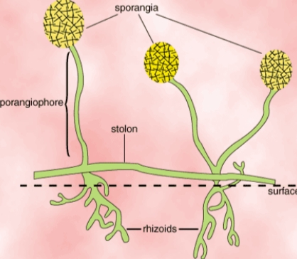
A rhizoid (Gr. rhiza = root + oeides = like) could also be a brief , root-like filamentous outgrowth of the thallus generally formed in tufts at rock bottom of small unicellular thalli or small porophores. Rhizoid is anchoring or attachment organ to the substratum and also as an organ of absorption of nutrients from substratum. Rhizoids are short, delicate filaments that contain protoplasm but no nuclei.
Rhizoids are common in lower fungi like Chytridiomycetes, Oomycetes and Zygomycetes. Some species produce a many-branched rhizomycelium. This is an in depth rhizoidal system that sometimes don't contains nuclei, but through which nuclei migrate. e.g. Cladochytrium sp. On rhizomycelium numerous sporangia develop. Such thalli are polycentric, that is, they form several reproductive centres rather than one one where the thallus is termed monocentric.
Appressorium (p1. appressorium; L. apprimere = to press against) could also be an easy or lobed structure of hyphal or germ tube and a pressing organ from which a flash infection peg usually grow and enter the epidermal cell of the host. It helps germ tube or hypha to connect to the surface of the host or substrates. These appressoria are formed from germ tubes of Uredinales (rust fungi), Erysiphales (powdery mildew fungi) and other fungi in their parasitic or saprophytic stages. In addition to giving anchorage, appressoria help the penetrating hyphae, branches to pierce the host cuticle. In fungi like Colletotrichum falcatum, germ tubes from conidia and resulting hyphae form appressoria on coming in touch with any pave like soil etc. These appressoria are thought to function as resting structures (chlamydospores) also.
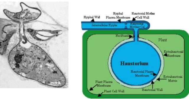
Haustoria (sing. haustorium; L. haustor = drinker) are special hyphal structures or outgrowths of somatic hyphae sent into the cell to soak up nutrients. The hyphal branch said to function as haustorium becomes extremely thin and pointed while piercing the hast cell membrane and expands within the cell cavity to make a wider, simple or branched haustorium. Haustoria could also be knob-like or balloon – like in shape, elongated or branched sort of a miniature rootage .
The hyphae of obligate parasites of plants like false mildew , mildew or rust fungi blight fungus etc ., produce haustoria. Hyphopodia: Hyphopodium (pl. hyphopodia Gr. hyphe = web + pous = foot) may be a small appendage with one or two cells long on an external hypha and performance as absorbing structures. The terminal cell of hyphopodium is expanded and rounded or pointed. Sometimes it produces a haustorium. e.g. Ectophytic fungi (Meliola aesariae) attacking leaves of green plants.
Mycelial strands are aggregates of parallel or interwoven undifferentiated hyphae, which adhere closely and are frequently anastomosed or cemented together. They are relatively loose (e.g. Sclerotium rolfsii growth on culture medium) compared to rhizomorph. They have no well-defined apical meristem. Mycelial strand formation is quite common in Basidiomycetes,
Ascomycetes and Deuteromycetes. Mycelial strands form the familiar 'spawn' of the cultivated mushroom, Agaricus bisporus. Mycelial strands are capable of translocating materials in both the directions. They are believed to afford means by which a fungus can extend a longtime food base and colonize a replacement substratum, by increasing the inoculum potential of the fungus at the point of colonization.
Rhizomorph is that the aggregation of highly differentiated hyphae with a well defined apical meristem, a central core of larger, thin walled, cells which are often darkly pigmented. These root-like aggregation is found within the honey mushroom or honey agaric honey mushroom (=Armillaria mellea). They grow faster than the mycelial strands. The growing tip of rhizomorph resembles that of a root tip. The fungus may spread underground from one rootage to a different by means of rhizomorph.
During certain stages of the life cycle of most fungi, the mycelium becomes organized into loosely or compactly woven tissues. These organized fungal tissues are called plectenchyma. There are two sorts of plectenchyma viz., prosenchyma and pseudoparenchyma. When the tissue is loosely woven and therefore the hyphae lie parallel to at least one another it's called prosenchyma. These tissues have distinguishable and typical elongated cells. Pseudoparenchyma consists of closely packed, more or less isodiametric or oval cells resembling the parenchyma cells of vascular plants. In this type of tissues hyphae lose their individuality and are not distinguishable. Prosenchyma cells are thin walled and cells in pseudoparenchyma.
Stromata and sclerotia are somatic structures of fungi.
A stroma is a compact, somatic structure or hyphal aggregation similar to a mattress or a cushion, on which or in which fructifications of fungi are usually formed. They may be of various shapes and sizes. Hyphal masses like acervuli, sporodochia, pionnotes etc. are the fertile stromata, which bear sporophores producing spores.
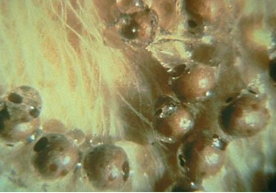
A sclerotium is a resting body formed by aggregation of somatic hyphae into dense, rounded, flattened, elongated or horn-shaped dark masses.
They are thick-walled resting structures, which contain food reserves. Sclerotia are hard structures resistant to unfavourable physical and chemical conditions. They may remain dormant for onger periods of time, sometimes for several years and germinate on the return of favourable conditions. The sclerotia on germination may be myceliogenous and produce directly the mycelium e.g. Sclerotium rolfsii, Rhizoctonia solani and S. cepivorum (white rot of onion).
They may be sporogenous and bear mass of spores. e.g. Botrytis cinerea. They may also be carpogenous where in they produce a spore fruit ( ascocarps or basidiocarps) bearing stalk. e.g. Sclerotinia sp. Claviceps purpurea (ergot of rye). Development of ascocarps is seen in Sclerotinia, where stalked cups or apothecia, bearing asci, arise from sclerotia. In Claviceps purpurea, sclerotia germinate and give rise to drumstick like structures called perithecial stromata, which contain perithecia, flask shaped cavities within which the asci are formed.
Mycorrhiza (pl. mycorrhizae; Gr. mykes = mushroom + rhiza = root) is the symbiotic association between higher plant roots and fungal mycelia. Many plants in nature have mycorrhizal associations. Mycorrhizal plants increase the surface area of the root system for better absorption of nutrients from soil especially when the soils are deficient in phosphorus.
The nature of association is believed to be symbiotic (mutualism), non-pathogenic or weakly pathogenic. There are three types of mycorrhizal fungal associations with plant roots.
They are ectotrophic or sheathing or ectomycorrhiza,. endotrophic or endomycorrhiza and ectendotrophicmycorrhiza.
The fungal nucleus has nuclear envelope comprising of two typical unit membrane and a central dense area referred to as nucleolus, which mainly contains RNA. In multinucleate hyphae, the nuclei may be interconnected by the endoplasmic reticulum. Vacuoles present inside the cell provide turgor needed for cell growth and maintenance of cell shape. Beside the osmotic function, they also store reserve materials. The chief storage products of fungi are glycogen and lipid. The apex of the hyphae are usually rich in vesicles and are called as apical vesicular complex (AVC) which helps within the transportation of products formed by the secretary action of golgi apparatus to the site where these products are utilized.
Specialized Somatic Structures Rhizoid
A rhizoid (Gr. rhiza = root + oeides = like) could also be a brief , root-like filamentous outgrowth of the thallus generally formed in tufts at rock bottom of small unicellular thalli or small porophores. Rhizoid is anchoring or attachment organ to the substratum and also as an organ of absorption of nutrients from substratum. Rhizoids are short, delicate filaments that contain protoplasm but no nuclei.
 |
| Source Wikipedia |
Rhizoids are common in lower fungi like Chytridiomycetes, Oomycetes and Zygomycetes. Some species produce a many-branched rhizomycelium. This is an in depth rhizoidal system that sometimes don't contains nuclei, but through which nuclei migrate. e.g. Cladochytrium sp. On rhizomycelium numerous sporangia develop. Such thalli are polycentric, that is, they form several reproductive centres rather than one one where the thallus is termed monocentric.
 |
| Source wikipedia |
Appressorium
Appressorium (p1. appressorium; L. apprimere = to press against) could also be an easy or lobed structure of hyphal or germ tube and a pressing organ from which a flash infection peg usually grow and enter the epidermal cell of the host. It helps germ tube or hypha to connect to the surface of the host or substrates. These appressoria are formed from germ tubes of Uredinales (rust fungi), Erysiphales (powdery mildew fungi) and other fungi in their parasitic or saprophytic stages. In addition to giving anchorage, appressoria help the penetrating hyphae, branches to pierce the host cuticle. In fungi like Colletotrichum falcatum, germ tubes from conidia and resulting hyphae form appressoria on coming in touch with any pave like soil etc. These appressoria are thought to function as resting structures (chlamydospores) also.
 |
| Source Wikipedia |
Haustoria
Haustoria (sing. haustorium; L. haustor = drinker) are special hyphal structures or outgrowths of somatic hyphae sent into the cell to soak up nutrients. The hyphal branch said to function as haustorium becomes extremely thin and pointed while piercing the hast cell membrane and expands within the cell cavity to make a wider, simple or branched haustorium. Haustoria could also be knob-like or balloon – like in shape, elongated or branched sort of a miniature rootage .
The hyphae of obligate parasites of plants like false mildew , mildew or rust fungi blight fungus etc ., produce haustoria. Hyphopodia: Hyphopodium (pl. hyphopodia Gr. hyphe = web + pous = foot) may be a small appendage with one or two cells long on an external hypha and performance as absorbing structures. The terminal cell of hyphopodium is expanded and rounded or pointed. Sometimes it produces a haustorium. e.g. Ectophytic fungi (Meliola aesariae) attacking leaves of green plants.
 |
| Source wikipedia |
Aggregations of hyphae and tissues
a. Mycelial strand
Mycelial strands are aggregates of parallel or interwoven undifferentiated hyphae, which adhere closely and are frequently anastomosed or cemented together. They are relatively loose (e.g. Sclerotium rolfsii growth on culture medium) compared to rhizomorph. They have no well-defined apical meristem. Mycelial strand formation is quite common in Basidiomycetes,
Ascomycetes and Deuteromycetes. Mycelial strands form the familiar 'spawn' of the cultivated mushroom, Agaricus bisporus. Mycelial strands are capable of translocating materials in both the directions. They are believed to afford means by which a fungus can extend a longtime food base and colonize a replacement substratum, by increasing the inoculum potential of the fungus at the point of colonization.
b. Rhizomorph
Rhizomorph is that the aggregation of highly differentiated hyphae with a well defined apical meristem, a central core of larger, thin walled, cells which are often darkly pigmented. These root-like aggregation is found within the honey mushroom or honey agaric honey mushroom (=Armillaria mellea). They grow faster than the mycelial strands. The growing tip of rhizomorph resembles that of a root tip. The fungus may spread underground from one rootage to a different by means of rhizomorph.
c. Fungal tissues
During certain stages of the life cycle of most fungi, the mycelium becomes organized into loosely or compactly woven tissues. These organized fungal tissues are called plectenchyma. There are two sorts of plectenchyma viz., prosenchyma and pseudoparenchyma. When the tissue is loosely woven and therefore the hyphae lie parallel to at least one another it's called prosenchyma. These tissues have distinguishable and typical elongated cells. Pseudoparenchyma consists of closely packed, more or less isodiametric or oval cells resembling the parenchyma cells of vascular plants. In this type of tissues hyphae lose their individuality and are not distinguishable. Prosenchyma cells are thin walled and cells in pseudoparenchyma.
Stroma and sclerotium
Stromata and sclerotia are somatic structures of fungi.
i. Stroma (pl. stromata; Gr. stroma = mattress)
A stroma is a compact, somatic structure or hyphal aggregation similar to a mattress or a cushion, on which or in which fructifications of fungi are usually formed. They may be of various shapes and sizes. Hyphal masses like acervuli, sporodochia, pionnotes etc. are the fertile stromata, which bear sporophores producing spores.
ii. Sclerotium (pl. sclerotia; Gr. skeleros =hard)
A sclerotium is a resting body formed by aggregation of somatic hyphae into dense, rounded, flattened, elongated or horn-shaped dark masses.
They are thick-walled resting structures, which contain food reserves. Sclerotia are hard structures resistant to unfavourable physical and chemical conditions. They may remain dormant for onger periods of time, sometimes for several years and germinate on the return of favourable conditions. The sclerotia on germination may be myceliogenous and produce directly the mycelium e.g. Sclerotium rolfsii, Rhizoctonia solani and S. cepivorum (white rot of onion).
 |
| Source Wikipedia |
They may be sporogenous and bear mass of spores. e.g. Botrytis cinerea. They may also be carpogenous where in they produce a spore fruit ( ascocarps or basidiocarps) bearing stalk. e.g. Sclerotinia sp. Claviceps purpurea (ergot of rye). Development of ascocarps is seen in Sclerotinia, where stalked cups or apothecia, bearing asci, arise from sclerotia. In Claviceps purpurea, sclerotia germinate and give rise to drumstick like structures called perithecial stromata, which contain perithecia, flask shaped cavities within which the asci are formed.
Mycorrhizae
Mycorrhiza (pl. mycorrhizae; Gr. mykes = mushroom + rhiza = root) is the symbiotic association between higher plant roots and fungal mycelia. Many plants in nature have mycorrhizal associations. Mycorrhizal plants increase the surface area of the root system for better absorption of nutrients from soil especially when the soils are deficient in phosphorus.
The nature of association is believed to be symbiotic (mutualism), non-pathogenic or weakly pathogenic. There are three types of mycorrhizal fungal associations with plant roots.
They are ectotrophic or sheathing or ectomycorrhiza,. endotrophic or endomycorrhiza and ectendotrophicmycorrhiza.







0 Comments
If you have any query let me know.