MUCOR - CLASSIFICATION, VEGETATIVE STRUCTURE, REPRODUCTION
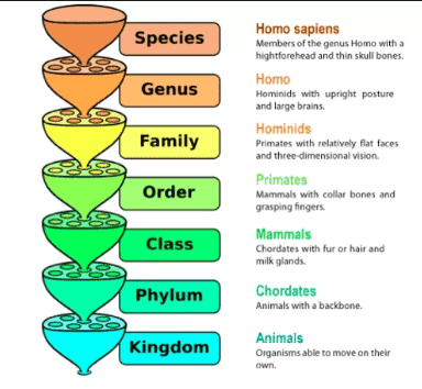
A.TAXONOMIC CLASSIFICATION
Division - Mycota
Sub-Division - Eumycotina
Class - Zygomycetes
Order - Mucorales
Family - Mucoraceae
Genus - Mucor
The genus mucor comprises about 42 species is a saprophytic fungus which is cosmopolitan in distribution. The species of mucor are found living on moist soil, dung and decaying plant and animal matter.
Mucor mucedo, is a common mould fungus growing sporophytically on various dacayed and rotten organic matter, moist leather, horse and cattle dung, etc. The species also spoils humans food like jam, jellies, bread, cheese etc. During rainy season, this fungus appears as white or grayish cottony mould on the substratum.
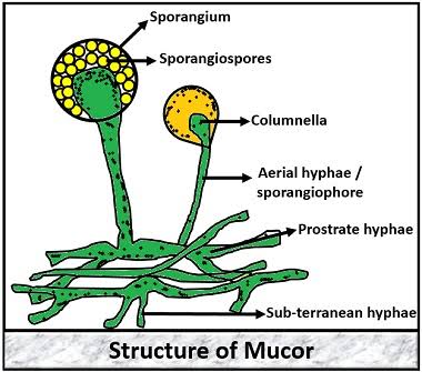
B. STRUCTURE OF THE VAGETATIVE BODY
The Vagetative body is stout and well develoved Mycelium which consists of a much branched filamentous coenocutic hyphae that spread in all directions over the substratum. Some of the hyphae penetrate deep into the substratum and serves as fixative and absorptive hyphae. Though, the hyphae are coenocytic, septa may appear later at the bases of the reproductive organ and occasionally on the older hyphae.
Cell wall of Mucor contains abundant chitosan, Besides, glucosamine, galactose, proteins, lipids, calcium etc are also present. The hyphae contain granular cytoplasm, large number if vacuoles, irregularly shaped numerous nuclei, droplets of oil and glycogen as reserve food.
C.REPRODUCTION
Mucor reproduce by both asexual or sexual process.
1.Asexual reproduction- Asexual reproduction takes place by the production of sporangiospore and clamydospore formation.
Sporangiospore formation- In this process, some of the unbranched hyphae, slightly broader then other grow up vertically in the air and are known as sporangiophores. The tip of sporangiophores swell up to farm a spherical sac-like structure called sporangia.
Through the sporangiophores, numerous nuclei along with cytoplasm flow into the sporangium and divide. The protoplasm collect densely in the peripheral region of the sporangium rather then the central region. Next, several small vacuoles forming a dome shaped foamy area appear within the protoplasm towards the base of the sporangial wall and is known as collumellaplasm. The collumellaplasm develop in collumella. The denser portion of the protoplasm with numerous nuclei constitutes the sporeplasm. The sporeplasm undergoes progressive cleavage, as a result of which develops into numerous non-flagellate sporangiospores.
Mature sporangiospores are thin walled, smooth, ovate and hyaline or coloured in Mass. At maturity, the sporangiospores are liberated by bursting of the sporangial wall and are disseminated by means of insects or wind. On reaching a suitable substratum, the sporangiospores germinates by producing one or more germ tube and ramify forming a new Mycelium.
Chlamydospore formation - These spore are formed when some of the hyphae break up by transverse wall into thick walled in chains. Later these cells round off and represent chlamydospores. Chlamydospore germinates to produce a new Mycelium.
Oidia formation - When species of mucor is allowed to grow in anaerobic culture medium especially in presence of CO2, a transverse breakage of hyphae takes place which result in the formation of Oidia.
2.Sexual Reproduction - Species of Mucor may be homothallic or hetrothallic. The sexual reproduction in Mucor is takes place by gametangial copulation methods. The copulating gametangia are isogamatangia, i.e., morphologically similiar gametes. In case of hetrothallic species, the two copulating isogamatangia arise from two physiological different and compatible hyphae of (+) and (-) strains. Numerous nuclei and cytoplasm flow into the tip of these hyphae which produces somewhat short and swollen lateral branches celled progametangia.
Two progametangia of opposite strains, i.e., (+) strain and (-) strains come in contact with each other through their apex - as a result the hyphae bearing progametangia are pushed apart. Now the tip of each progametangium is cut off by transverse wall dividing the progametangium into a distil gametangium and basal suspensor cell. In the meantime, the common wall at the point of contact between the two gametangia break down and nuclei of (+) and (-) strains fuse to from diploid nuclei. As a result multinucleate zygospore is formed. The diploid nuclei of zygospore undergo meiotic divisions and hence, the zygospore contains haploid nuclei. The zygospore is set free by disintegration of the parent Mycelia.
After a period of rest over a month, zygospore germinates by producing a long germ tube called promycelium. The tip of promycelium develops a typical sporangium called zygosprangium or germ sporangium. Inside the zygosporangium, one kind of spore, either (+) or (-) strain spore are produced. Each of the spore after the liberation of zygosporangium germinates by producing a germ tube to give rise to new Mycelium.
Sometime gametangia fails to fuse and then a single gametangium may directly develop into a thick walled structure called azygospore or parthenospore. Azygospore directly germinates into new Mycelium in due course.
 |
| Source Wikipedia |
C.REPRODUCTION
Mucor reproduce by both asexual or sexual process.
1.Asexual reproduction- Asexual reproduction takes place by the production of sporangiospore and clamydospore formation.
Sporangiospore formation- In this process, some of the unbranched hyphae, slightly broader then other grow up vertically in the air and are known as sporangiophores. The tip of sporangiophores swell up to farm a spherical sac-like structure called sporangia.
Through the sporangiophores, numerous nuclei along with cytoplasm flow into the sporangium and divide. The protoplasm collect densely in the peripheral region of the sporangium rather then the central region. Next, several small vacuoles forming a dome shaped foamy area appear within the protoplasm towards the base of the sporangial wall and is known as collumellaplasm. The collumellaplasm develop in collumella. The denser portion of the protoplasm with numerous nuclei constitutes the sporeplasm. The sporeplasm undergoes progressive cleavage, as a result of which develops into numerous non-flagellate sporangiospores.
Mature sporangiospores are thin walled, smooth, ovate and hyaline or coloured in Mass. At maturity, the sporangiospores are liberated by bursting of the sporangial wall and are disseminated by means of insects or wind. On reaching a suitable substratum, the sporangiospores germinates by producing one or more germ tube and ramify forming a new Mycelium.
Chlamydospore formation - These spore are formed when some of the hyphae break up by transverse wall into thick walled in chains. Later these cells round off and represent chlamydospores. Chlamydospore germinates to produce a new Mycelium.
Oidia formation - When species of mucor is allowed to grow in anaerobic culture medium especially in presence of CO2, a transverse breakage of hyphae takes place which result in the formation of Oidia.
 |
| Source Wikipedia |
2.Sexual Reproduction - Species of Mucor may be homothallic or hetrothallic. The sexual reproduction in Mucor is takes place by gametangial copulation methods. The copulating gametangia are isogamatangia, i.e., morphologically similiar gametes. In case of hetrothallic species, the two copulating isogamatangia arise from two physiological different and compatible hyphae of (+) and (-) strains. Numerous nuclei and cytoplasm flow into the tip of these hyphae which produces somewhat short and swollen lateral branches celled progametangia.
Two progametangia of opposite strains, i.e., (+) strain and (-) strains come in contact with each other through their apex - as a result the hyphae bearing progametangia are pushed apart. Now the tip of each progametangium is cut off by transverse wall dividing the progametangium into a distil gametangium and basal suspensor cell. In the meantime, the common wall at the point of contact between the two gametangia break down and nuclei of (+) and (-) strains fuse to from diploid nuclei. As a result multinucleate zygospore is formed. The diploid nuclei of zygospore undergo meiotic divisions and hence, the zygospore contains haploid nuclei. The zygospore is set free by disintegration of the parent Mycelia.
After a period of rest over a month, zygospore germinates by producing a long germ tube called promycelium. The tip of promycelium develops a typical sporangium called zygosprangium or germ sporangium. Inside the zygosporangium, one kind of spore, either (+) or (-) strain spore are produced. Each of the spore after the liberation of zygosporangium germinates by producing a germ tube to give rise to new Mycelium.
Sometime gametangia fails to fuse and then a single gametangium may directly develop into a thick walled structure called azygospore or parthenospore. Azygospore directly germinates into new Mycelium in due course.
 |
| Source Wikipedia |


0 Comments
If you have any query let me know.