A. Classification
The genus Selaginella is commonly known as club moss or spike moss. Selaginella is a large genus comprising about 700 species and is world wide in distribution. Some species of Selaginella are found to grow in template regions but majority of them occur in the rain forest of tropical countries.
The species of Selaginella is found to grow on the ground, on damp shaded, and humid condition. Some of the species are occur in arid regions of the world. Temprate species are found to grow on damp shaded side of the hills.
About 55 species are found to occur in India. Of these common species are S.rupestries, S.pentagona, S.megaphylla etc.
B. Structure of the sporophyte
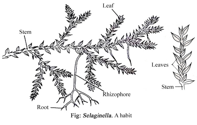
1. External Structure:- The sporophyte i.e., plant body is well differentiated into - stem, roots and leaves-
Stem- The stem is long , slender , usually dosiventral and prostrste with erect branches. In some species the stem is erect. The stem may be un-branched or dichotomously branched. From each ramification of the stem, colourless, leafless, elongated and cylindrical appendages known as rhizophores develop. The rhizophores develop downwardly into the soil and give rise to small tuft adventious roots at their free ends.
Leaves- Stems and branches bear numerous small, lanceolate, ovate to filiform leaves which are aaranged in spiral, decussate pair of four longitudinal rows.
Leaves are generally thin and delicate in texture, and are provided with unbranched mid vein.
Roots- First root i.e., primary root is short lived, later delicate adventious root arise from the underside of the stem and from the tip rhizophores also. Roots are delicate and branching is dichotomous.
2. Internal Structure:-
a) T.S of stem: The internal structure of aerial shoot shows the following tissue systems-
Epidermis- Epidermis is one cell in thickness and it consists of parenchymatous cell. Stomata are absent in the epidermal layer.
Cortex- Cortex is thick and composed entirely of thin-walled, green, parenchymatous cells without intercellular spaces, or partly sclerenchymatous cells farming hypodermis and parenchymatous cells. True endodermis is absent , instead endosermal cell modified into radially elongated cells known as tuberculae, by means of which Stele or Steles are attached to the cortex. The tuberculae cells contain casparian strips.
Stele- The stelar organization is varies in different species. The Stele is protostelic in nature with exarch xylem , the number of which varies from one (monostelic) to several i.e., 1,2,3,4, etc (polystelic). Each Stele is limited externally by a layer of pericycle.
b) T.S of Root: The root in cross section shows one cell layer thick Epidermis. Cortex is like that of stem but is provided with endosermis. Stele is protostelic, which is monarch and exarch.
c) T.S of Leaves: The leaf in cross section shows a distinct upper and lower Epidermis , each of one celled in thickness, and undifferentiated mesophyll tissue is composed of more or less elongated and similiar cells with intercellular spaces. Vascular bundle is concentric. Phloem surround the xylem.
C) Reproduction
Sporophyte of Selaginella reproduce both by vegetative means by production of spores.
1. Vegetative reproduction- vegetative reproduction takes place by following methods-
a) Fragmentation- it is affected only in species that grow under humid condition. In this case the trailing branches of the stem develop adventious branches and later get disconnected from their parent plant and grow into separate individual plants.
b) Tubers: Formation of tubers have been reported in S.abyssinica. The tubers bears rudimentary scales and appear towards the end of the growing season at the tip of the of the underground branches that arise from base of the stem. During unfavorable condition the aerial part of the plant die and the tubers enable the plant to perrenate. At the Advent of favourable condition the tubers germinate to produce new plant.
c) Resting buds: Resting buds have been report to develop at the ends of some aerial branches in S.chrysocaulos. It is formed as result of compact aarangement of the leaves . The buds give off rhizophores that bears roots at their tips and fix them to the soil. The resting buds survive the unfavorable periods when the rest of the plant dies. They grow into new individuals at the return of the favourable condition.
2. Spore Formation- In Selaginella, the spores are formed in a specialized reproductive structure known as strobili or cone.
The cone i.e., strobillus varies in size from 5mm to 7cm. They are cylendrical or quariangular and are borne at the apices of the main stem or on lateral branches. Each strobillus consists of an axis upon which two types of sporophylls are arranged spirally. The megasporophylls appear at the base and microsporophylls at the above portion of the strobillus.
Each megasporophylls bears a single stalked megasporangium in it's axil on it's upper side. Similiarly microsporophylls bears a single microsporangium in it's axil on it's upper side. Megasporangia are larger in size than the microsporangia. Both the types of sporangia are provided with a jacket wall of sterile cells of two- celled thickness. Within the jacket wall lies the sporogenous tissue , which is surrounded externally by a prominent layer of nutritive tissue known as tapetum.
Sporogenous tissue of each megasporangium differentiates into megaspores mother cells , but all of them except one degenerates. The survinving megaspores mother cells by reduction divisions give rise to four megaspores only one or two survive , other degenerates.
Within the microsporangium, sporogenous tissues later on differentiates into Microspore mother cells , all of which except of very few , by reduction divisions give rise to spore- tetrads. Thus, each microsporangium contains numerous Microspore.
D. Structure of the Gametophyte
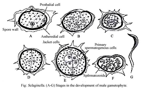
1.Male Gametophyte : Microspore is the first cell of male Gametophyte. Each Microspore is small, spherico-tetrahedral and provided with two coats, viz, outer thick ornamental exine and inner thick delicate intine.
Germination of Microspore takes place within the microsporangium. Microspore nucleus first divides to form small lense shaped prothallial cell at one side and the larger anthredial initial. The prothallial cell divides no further but the anthredial initial divided and re-divides farming 12-celled structure , the so called antheridium. Now the male Gametophyte consists of 13 cells, the , of these 13 cells , the central four cells constitute the primary spermatogenous cells, the eight cells surrounding the primary spermatogenous cells constitute the sterile jacket cells.
The primary spermatogenous cells divide and re-divide forming 128-256 sperm mother cell is then metamorphosed into biflagellate sperms.
2. Female Gametophyte : megaspores is the first cell of female Gametophyte. Megaspores are larger and tetrahedral in shape and consists of outer sculpture thick exine and inner thin intine.
The female Gametophyte also begins to germinates while the megaspores is still within the megasporangium. The germinating megaspores first enlarge in size and now consists of three wall layer and a thin layer of peripheral cytoplasm enclosing the nucleus .
3. Fertilization: It may takes place while the female Gametophyte is still within the megasporangium or after the megasporangium has fallen to the ground.
The sperm after liberation swim to the archegonia in dew or in rain water and of them ultimately fertilize the egg. As a result (2n) zygote is formed, with the formation of zygote , diploid spot generation begins.
Divisions-Lycophyta
Class-Lycopsida
Order-Selaginellales
Famy-Selaginellaceae
Genus-Selaginella
The species of Selaginella is found to grow on the ground, on damp shaded, and humid condition. Some of the species are occur in arid regions of the world. Temprate species are found to grow on damp shaded side of the hills.
About 55 species are found to occur in India. Of these common species are S.rupestries, S.pentagona, S.megaphylla etc.
B. Structure of the sporophyte
1. External Structure:- The sporophyte i.e., plant body is well differentiated into - stem, roots and leaves-
 |
| Source Wikipedia |
Stem- The stem is long , slender , usually dosiventral and prostrste with erect branches. In some species the stem is erect. The stem may be un-branched or dichotomously branched. From each ramification of the stem, colourless, leafless, elongated and cylindrical appendages known as rhizophores develop. The rhizophores develop downwardly into the soil and give rise to small tuft adventious roots at their free ends.
Leaves- Stems and branches bear numerous small, lanceolate, ovate to filiform leaves which are aaranged in spiral, decussate pair of four longitudinal rows.
Leaves are generally thin and delicate in texture, and are provided with unbranched mid vein.
Roots- First root i.e., primary root is short lived, later delicate adventious root arise from the underside of the stem and from the tip rhizophores also. Roots are delicate and branching is dichotomous.
2. Internal Structure:-
a) T.S of stem: The internal structure of aerial shoot shows the following tissue systems-
Epidermis- Epidermis is one cell in thickness and it consists of parenchymatous cell. Stomata are absent in the epidermal layer.
Cortex- Cortex is thick and composed entirely of thin-walled, green, parenchymatous cells without intercellular spaces, or partly sclerenchymatous cells farming hypodermis and parenchymatous cells. True endodermis is absent , instead endosermal cell modified into radially elongated cells known as tuberculae, by means of which Stele or Steles are attached to the cortex. The tuberculae cells contain casparian strips.
Stele- The stelar organization is varies in different species. The Stele is protostelic in nature with exarch xylem , the number of which varies from one (monostelic) to several i.e., 1,2,3,4, etc (polystelic). Each Stele is limited externally by a layer of pericycle.
b) T.S of Root: The root in cross section shows one cell layer thick Epidermis. Cortex is like that of stem but is provided with endosermis. Stele is protostelic, which is monarch and exarch.
c) T.S of Leaves: The leaf in cross section shows a distinct upper and lower Epidermis , each of one celled in thickness, and undifferentiated mesophyll tissue is composed of more or less elongated and similiar cells with intercellular spaces. Vascular bundle is concentric. Phloem surround the xylem.
 |
| Source Wikipedia |
C) Reproduction
Sporophyte of Selaginella reproduce both by vegetative means by production of spores.
1. Vegetative reproduction- vegetative reproduction takes place by following methods-
a) Fragmentation- it is affected only in species that grow under humid condition. In this case the trailing branches of the stem develop adventious branches and later get disconnected from their parent plant and grow into separate individual plants.
b) Tubers: Formation of tubers have been reported in S.abyssinica. The tubers bears rudimentary scales and appear towards the end of the growing season at the tip of the of the underground branches that arise from base of the stem. During unfavorable condition the aerial part of the plant die and the tubers enable the plant to perrenate. At the Advent of favourable condition the tubers germinate to produce new plant.
c) Resting buds: Resting buds have been report to develop at the ends of some aerial branches in S.chrysocaulos. It is formed as result of compact aarangement of the leaves . The buds give off rhizophores that bears roots at their tips and fix them to the soil. The resting buds survive the unfavorable periods when the rest of the plant dies. They grow into new individuals at the return of the favourable condition.
2. Spore Formation- In Selaginella, the spores are formed in a specialized reproductive structure known as strobili or cone.
The cone i.e., strobillus varies in size from 5mm to 7cm. They are cylendrical or quariangular and are borne at the apices of the main stem or on lateral branches. Each strobillus consists of an axis upon which two types of sporophylls are arranged spirally. The megasporophylls appear at the base and microsporophylls at the above portion of the strobillus.
Each megasporophylls bears a single stalked megasporangium in it's axil on it's upper side. Similiarly microsporophylls bears a single microsporangium in it's axil on it's upper side. Megasporangia are larger in size than the microsporangia. Both the types of sporangia are provided with a jacket wall of sterile cells of two- celled thickness. Within the jacket wall lies the sporogenous tissue , which is surrounded externally by a prominent layer of nutritive tissue known as tapetum.
Sporogenous tissue of each megasporangium differentiates into megaspores mother cells , but all of them except one degenerates. The survinving megaspores mother cells by reduction divisions give rise to four megaspores only one or two survive , other degenerates.
Within the microsporangium, sporogenous tissues later on differentiates into Microspore mother cells , all of which except of very few , by reduction divisions give rise to spore- tetrads. Thus, each microsporangium contains numerous Microspore.
D. Structure of the Gametophyte
1.Male Gametophyte : Microspore is the first cell of male Gametophyte. Each Microspore is small, spherico-tetrahedral and provided with two coats, viz, outer thick ornamental exine and inner thick delicate intine.
Germination of Microspore takes place within the microsporangium. Microspore nucleus first divides to form small lense shaped prothallial cell at one side and the larger anthredial initial. The prothallial cell divides no further but the anthredial initial divided and re-divides farming 12-celled structure , the so called antheridium. Now the male Gametophyte consists of 13 cells, the , of these 13 cells , the central four cells constitute the primary spermatogenous cells, the eight cells surrounding the primary spermatogenous cells constitute the sterile jacket cells.
The primary spermatogenous cells divide and re-divide forming 128-256 sperm mother cell is then metamorphosed into biflagellate sperms.
 |
| Source Wikipedia |
The female Gametophyte also begins to germinates while the megaspores is still within the megasporangium. The germinating megaspores first enlarge in size and now consists of three wall layer and a thin layer of peripheral cytoplasm enclosing the nucleus .
 |
| Source Wikipedia |
The sperm after liberation swim to the archegonia in dew or in rain water and of them ultimately fertilize the egg. As a result (2n) zygote is formed, with the formation of zygote , diploid spot generation begins.






0 Comments
If you have any query let me know.